Translational Imaging Research Facility
3T MRI Core Facility
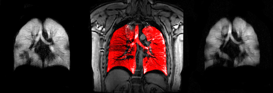
Research at the 3T MRI Core Facility is focused on in-vivo studies of physiology and function using a variety of advanced magnetic resonance imaging methods. Major interests include cancer, cardiovascular, maternal health, neuro-degenerative, orthopedics and respiratory disease, which are studied in both humans and animal models.
The 3T MRI Core Facility is a unique regional resource for biomedical research possessing a state-of-the-art clinical 3.0 Tesla MRI scanner, insertable high-performance gradient hardware for micro-imaging of pre-clinical models, polarization technology for gases and liquids and relaxometry infrastructure for characterization of MR contrast agents.
The facility resources are available to peer-reviewed grant funded scientific collaborators with appropriate Research Ethics Board (REB) protocols in place.
Some of the current studies include Alzheimer's studies, carotid artery imaging, cellular and molecular imaging with novel contrast media, knee cartilage imaging, advanced prostate cancer imaging and pulmonary imaging of lung diseases with hyperpolarized gases.
See: Robarts Research Institute 3T MRI Core Facility
Major Facility Infrastructure
3T MRI
Central to this facility is a General Electric Healthcare Discovery MR750 3.0T™ clinical magnetic resonance imaging scanner with a full complement of anatomy-specific RF coils for human imaging. This imaging platform has been upgraded to include broadband RF hardware capable of imaging hyperpolarized gases (3He & 129He) and liquids (13C) as contrast agents as well as iron or 19F-labeled labeled cells. The scanner has also been equipped with an insertable high-performance gradient system and compatible RF hardware for micro-imaging of small animals using a variety of clinically relevant imaging protocols.
Dynamic Nuclear Polarization Laboratory
The Dynamic Nuclear Polarization Laboratory contains an Oxford Instruments HyperSense™ polarizer for in vitro magnetization of 13C-enriched liquids. These simple endogenous molecules are subsequently injected as real-time probes of regional in vivo metabolism in animal models of disease. Researchers are using this technology to probe glycolysis, pH and necrosis in tumour models, assess radiation-induced lung inflammation and fetal health.
Relaxometry Laboratory
This laboratory contains fast field-cycling relaxometry hardware for measuring the nuclear magnetic relaxation dispersion of novel paramagnetic contrast agents. A stand-alone, Stelar Spinmaster™ FFC2000 1T C/DC relaxometer is capable of acquiring automated temperature-controlled measurements of spin-lattice relaxation from the earth’s field up to 1 Tesla. A second insertable Stelar relaxometer prototype is capable of making these measurements ± 0.25T from the clinical field strength in which the system is inserted. Collaborations with contrast agent chemistry groups to characterize new MRI agents are encouraged.
3T MRI Scientific Directors
Dr. Paula Foster - pfoster@robarts.ca
Dr. Timothy Scholl - tscholl@robarts.ca
Visit Translational Imaging Research Facility website for more information about the 3T MRI Core Facility, including booking instructions and on-line schedule. Billing information for 3T MRI Core Facility access can be found under ‘SOP Documents’.
For more information on the facility, booking etc. please contact the facility staff.
MR Technologist
D. Reese
519-931-5777 ext. 24109
E-mail: dreese@robarts.ca
MR Physicist
T. Wade
519-931-5777 ext. 24243
E-mail: twade@robarts.ca
Centre for Translational Radiographic Imaging
The Centre for Translational Radiographic Imaging (CenTRI) within the Translational Imaging research Facility at Western’s Robarts Research Institute is a platform for the development and application of state-of-the-art radiographic imaging equipment. Operating as a close collaboration between imaging scientists, physicians, and industry partners, the Centre for Translational Radiographic Imaging bridges the gap between development of novel technology and routine clinical application. Developments withing the Centre for Translation Radiographic Imaging enhance the outcomes of patients with neurological, cardiac, pulmonary, oncological, and musculoskeletal diseases.
The facility resources are available to peer-reviewed grant funded scientific collaborators with appropriate Research Ethics Board (REB) protocols in place.
Some of the current radiographic studies include brain perfusion, dynamic cardiac imaging, lung imaging, and orthopaedic implant migration.
See: Robarts Research Institute Translational Imaging Research Facility
Major Facility Infrastructure
CT Scanner
This facility has a Canon Acquillion ONE PRISM Edition CT scanner with state-of-the-art AI-assisted technologies that produce volumetric reconstructions with low noise, fine image texture, sharp high contrast resolution, and clear low contrast detectability. This imaging platform includes a fully integrated end-to-end spectral imaging workflow which harnesses the temporal benefits of rapid kV switching combined with a Deep Learning reconstruction algorithm that delivers virtual monochromatic images and iodine mapping. The scanner has also been equipped with a PUREVision CT detector, capable a 500-micron isotropic resolution images using either conventional, iterative, or deep learning reconstruction algorithms.
Angiography System
The Canon Alphenix Core+ Is a five-axis floor-mounted C-arm that allows for head-to-toe and fingertip-to-fingertip coverage required to perform various interventional procedures. This imaging system has been integrated with real-time dose tracking, parametric imaging, needle guidance, and multi-modality fusion. In addition to conventional radiographic and fluoroscopic imaging, this imaging system is capable of volumetric acquisitions with 306-micron isotropic resolution.
Digital Tomosynthesis System
The Carestream DRX Evolution is a state-of-the-art general x-ray suite equipped with a wireless DRX Plus 3543C, motorized overhead tube and wallstand, and a versatile table. The imaging system can acquire digital tomosynthesis images that are analyzed to provide depth information about patient anatomy by separating overlying structures.
Weight-Bearing CT Scanner
Carestream OnSight 3D Extremity Scanner is a cone-beam CT scanner equipped three X-ray sources that utilizes iterative reconstruction, advanced scatter, and metal artifact-correction algorithms to deliver high resolution 3D images with a 260-micron isotropic resolution. This system has a motorized gantry allowing for a variety of positions for the extremities and enabling weight-bearing positions to assess the lower extremities under a natural load.
Radiostereometry Laboratory
This laboratory contains two GE Proteus XR/a systems, used in conjunction with a calibration cage, to assess micromotion in orthopaedic implants for up to two years following surgery. Through the use of specialized software, joint kinematics and orthopaedic implant migration can be detected within 50-microns.
CenTRI Scientific Directors
Dr. Matthew Teeter - mteeter@robarts.ca
Dr. David Holdsworth - dholdsworth@robarts.ca
Visit Translational Imaging Research Facility website for more information about CenTRI, including booking instructions and on-line schedule. Billing information for CenTRI access can be found under ‘SOP Documents’.
For more information on the facility, booking etc. please contact the facility staff.
CenTRI Facility Technologist
B. Sinclair
519-931-5777 ext. 24065
E-mail: bsincl7@uwo.ca
Radiation Safety Officer
Fadi Al Jallad
519-931-5777 ext. 84821
E-mail: faljalla@uwo.ca


