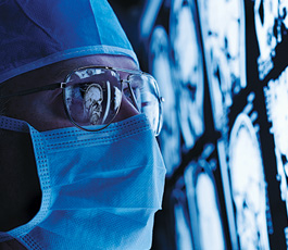
Translational Imaging Research Facility
The Translational Imaging Research Facility (TIRF) represents an unparalleled resource for state-of-the-art MR and X-ray imaging research. Major infrastructure includes: a multi-nuclear-capable clinical MRI system (GE 3T MR750) with an insertable high-performance gradient system for micro-imaging of preclinical models, a state-of-the-art CT scanner (Canon Acquilion ONE PRISM Edition), an angiography system (Canon Alphenix Core+), a digital tomosynthesis system (CareStream DRX-Evolution Plus), a weight-bearing cone-beam CT scanner (CareStream OnSight 3D Extremity System), and a radiostereometry laboratory (GE Proteus XR/a, Carestream DRX, & Fujifilm Capsula). The facility is a valuable resource for x-ray based, multi-nuclear (non-hydrogen), conventional (hydrogen), and other advanced MR imaging methods.
Clinical MRI Research
The main focus of clinical research includes multi-nuclear MR imaging of patients with lung diseases using hyperpolarized (3He & 129Xe) gases, as well as, evaluation of endogenous 23Na storage in human tissue of patients with kidney disease. Other supported clinical research projects at the TIRF focus on neuro-microvascular and neurodegenerative diseases, neuropsychiatric disorders, MSK disorders, liver disease, and several industry sponsored clinical trials.
Non-Clinical MRI Research
Non-clinical MR imaging studies include cellular and molecular imaging of tumours with novel contrast media (19F, 13C, and iron oxide nanoparticles) and development of MRI reporter gene systems for tracking cell and immunotherapies.
Clinical X-ray Research
There is a broad spectrum of clinical research that includes dynamic CT scans, contrast-enhanced cardiac imaging, and assessment of lung function following COVID-19 infections. Other x-ray based clinical research includes several industry sponsored studies to assess orthopaedic implant migration and surgical approaches.
Non-Clinical X-ray Research
Non-clinical X-ray imaging studies include quantitative assessment using image quality phantoms and development of tools to assess brain and cardiac perfusion.
Please email mrbookings@robarts.ca to schedule time for your 3 Tesla MRI studies, or centri@uwo.ca to schedule time for your x-ray studies.
CONTACT US
3T MRI Research Facility
Robarts Research Institute
Western University
1151 Richmond Street
London, Ontario Canada N6A 3K7
t. 519.931.5777
e. TIRFmri@uwo.ca
Centre for Translational Radiographic Imaging
Robarts Research Institute
Western University
1151 Richmond Street
London, Ontario Canada N6A 3K7
t. 519.931.5777
e. centri@uwo.ca


