Core Scientists
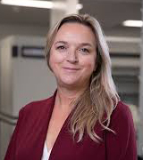
Paula Foster PhD
Co-Director/Research ScientistAreas of Expertise: Cellular imaging using iron-labeled cells and MRI, imaging tissue inflammation, traumatic spinal cord injury, tissue histology
Biography: As the leader of the Cellular and Molecular Imaging program at Robarts, Dr. Foster is developing imaging and cell labelling technology that uses ultra-high resolution MRI to detect cells labeled with magnetic nanoparticles. This advanced MRI technology has enormous potential, with limitless applications in a number of diseases and disorders. The Foster lab is currently focused on the use of these techniques to track stem cells used for tissue repair and regeneration and to monitor cancer cell metastasis and immune cells used as cancer treatments.
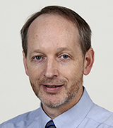
David Holdsworth PhD
Co-Director/Research ScientistAreas of Expertise: Quantitative imaging, dynamic imaging, computed tomography, musculoskeletal MRI, image-based finite-element models, musculoskeletal biomechanics, wireless telemetry, image-based design, 3D-printing
Biography: Dr. David Holdsworth is a Scientist in the Imaging group at the Robarts Research Institute. He is also a Professor in the Departments of Surgery and Medical Biophysics in the Schulich School of Medicine and Dentistry, at the University of Western Ontario. For most of the past 15 years, Dr. Holdsworth has been involved in the development of vascular imaging systems, for use in stroke diagnosis and therapy. In 2007 Dr. Holdsworth became the Dr. Sandy Kirkley Chair in Musculoskeletal Research and has shifted the focus of his research to musculoskeletal disease, with projects ranging from basic skeletal research to clinical therapy. Dr. Holdsworth and his team have developed new methods for musculoskeletal disease detection and treatment for both basic pre-clinical and clinical applications. With collaborators in surgery and engineering, he is developing new techniques to image the interface between bones and metal implants, and to improve techniques for radiostereometric analysis following joint replacement.

Charlie McKenzie PhD
Research ScientistAreas of Expertise: Magnetic Resonance Imaging (MRI) of pregnancy: development of coil arrays, pulse sequences and image reconstructions; correction of motion; Metabolic MRI of the fetus and placenta.
Biography: Research in the McKenzie Lab focuses on the development of new MRI image acquisition and reconstruction techniques, with a particular focus on MRI during pregnancy. We are developing new methods for direct imaging of fetal and placental metabolism, in addition to techniques for detecting the consequences of metabolic dysfunction during pregnancy. Trainee's in the lab come from a wide variety of basic and applied science disciplines, including biophysics, physics, computer science, biochemistry, physiology and biomedical engineering. Work in the lab occurs in both research and clinical settings around London, including the main Western campus, the Robarts Research Institute, the Children's Health Research Institute and London Health Sciences Centre

Grace Parraga PhD
Research ScientistAreas of Expertise: COPD, Asthma, Cystic Fibrosis, Lung Cancer, respiratory system imaging
Biography: Dr. Parraga's lab is fascinated by the interactions of lung growth and development with lung aging and disease. The lungs are central to homeostasis and to life so that without optimal lung structure and function, all other physiological systems in humans are compromised and eventually fail. Lung abnormalities are expressed “silently” until disease mechanisms are well-established or irreversible which makes detection and a deep understanding of lung disease and its mechanisms extremely challenging. Research in our laboratory is directed towards a detailed understanding of lung structure and function, with an emphasis on the development of human medical imaging tools.
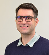
John Ronald PhD
Research ScientistAreas of Expertise: Cancer, Molecular Imaging, Reporter Genes, Cell Tracking, In Vitro Diagnostics
Biography: As we enter this era of more personalized and precise medicine, new technologies are needed that can sensitively, accurately and non-invasively detect molecular activities within the body over the course of an individual’s entire life. My lab’s research focuses on pioneering novel molecular and cellular imaging technologies that will hopefully meet these needs. We have a particular interest on improved early cancer detection, as well as improved monitoring of state-of-the-art gene-based and cell-based therapies for cancer and other diseases. To accomplish this we are investigating the development of novel molecular biology platforms that strategically integrates disease-specific activatable expression systems with both biofluid-based and multimodality imaging readouts. This work is at the interface of molecular and cell biology, imaging sciences, and nanomedicine and requires a multidisciplinary approach to devise innovative solutions to some of today’s most difficult biomedical problems.

Timothy Scholl PhD
Co-Director/Research ScientistAreas of Expertise: Hyperpolarized probes for metabolic imaging such as for characterizing and staging a tumor, and early stage treatment
Biography: My research program at the Robarts Research Institute is focused on the development of novel molecular probes for magnetic resonance imaging. Molecular imaging is a non-invasive tool for visualization of cellular function in living organisms. For magnetic resonance imaging (MRI), cellular function is imaged by injecting a contrast agent into an organism that produces quantifiable changes in the image reflecting variations in metabolic function. These compounds may be designed to bind preferentially to a specific protein or be metabolized directly by cells so that the effects of their bio-distribution can be imaged using MRI. Compared with other imaging modalities, the intrinsic sensitivity of MRI is low and the challenge is to produce newer contrast agents that can be effective at lower concentrations. The chief property affecting sensitivity is the contrast agent “relaxivity” – the effectiveness of an agent to cause signal enhancement through increased magnetic relaxation of nearby tissue. My laboratory is set up to measure relaxivity of contrast agents using dedicated field-cycling relaxometers. I am also interested in the application of a new type of contrast agent based on hyperpolarized 13C-enriched endogenous compounds. These slowly relaxing agents are highly magnetized (hyperpolarized) in vitro and then are injected into an animal model and spectroscopically imaged along with their metabolic products. The spatial distributions of the metabolites yield valuable biomarkers about in vivo cellular processes that can be used to detect and grade tumors and gauge their response to therapy. The immediate objectives of my research program are three-fold: 1) application of hyperpolarized compounds as contrast agents to probe altered cellular metabolism in tumors; 2) development of our dreMR technology and its translation to in vivo imaging with animal models; 3) measurement of relaxivity profiles of novel contrast agents.
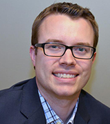
Matthew Teether PhD
Co-Director/Research ScientistAreas of Expertise: Additive Manufacturing, Arthritis, Bioengineering, Biomechanics, Clinical Outcomes, Clinical Trials, Implantable & Wearable Devices, Musculoskeletal Imaging, Orthopaedic Surgery
Biography: Dr. Matthew Teeter leads the basic science research program for the Joint Replacement Institute at London Health Sciences Centre, University Hospital. He is a Scientist at the Lawson Health Research Institute, Scientist at the Robarts Research Institute, and an Assistant Professor in the Departments of Surgery and of Medical Biophysics, Schulich School of Medicine & Dentistry at Western University. Dr. Teeter’s research focuses on image-based orthopedic implant design and evaluation, including the use of wearable sensors, 3D printing technologies, and imaging techniques such as micro-CT, CT, RSA, and single-plane fluoroscopy. He also directs the Implant Retrieval Laboratory at LHSC. Dr. Teeter received his BSc in Biomedical Science from the University of Guelph and his PhD in Medical Biophysics from Western University, where he was a Graduate Fellow in Musculoskeletal Health Research and Leadership as part of the CIHR Joint Motion Program. He received national awards at all levels of his graduate and fellowship training from the CIHR. He has been recognized with the Mark Coventry Award from the Knee Society, the John Charles Polanyi Prize in Physiology or Medicine, an Ontario Early Researcher Award, and a CIHR New Investigator Award.


