Services
This facility is can provide histology services or provide training for research staff. Please contact the lab for pricing information.
Equipment
Nanostring GeoMx - Digital Spatial Profiler with a flexible and robust spatial multiomic platform for analysis of FFPE and fresh frozen tissue sections.
Nanostring CosMx - Spatial Molecular Imager System with a high-plex spatial multiomic single cell imaging platform.
AS-410M Dainippon Seiki Automated Tissue Sectioner - Large capacity paraffin tissue sectioner for up to 800 sequential tissue sections for histology and 3D reconstruction.
Leica Aperio AT2 Brightfield Slide Scanner - Scans brightfield images at 5x, 20x or 40x magnification and has an image analysis workstation and algorithms.
Leica Versa Fluorescent Slide Scanner - Fluorescent slide scanner with 1.25x, 5x, 10x, 20x, 40x and 60x magnification and 8 fluorescent filter cubes.
3D Histech Tissue Microarray Master - Automated Tissue Microarray with 0.6, 1.0. 1.5 and 2 mm cores.
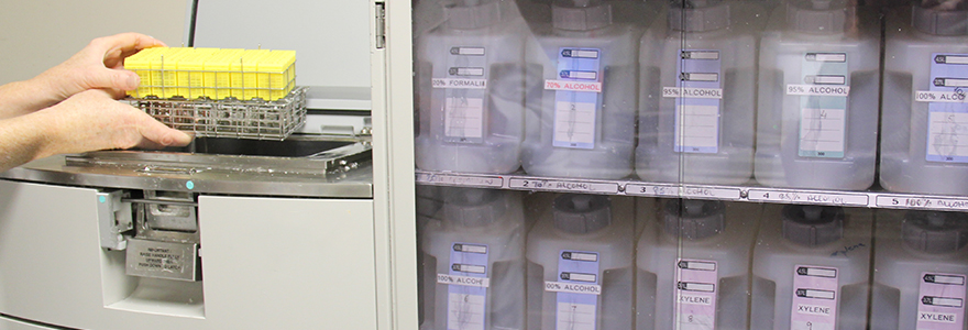
Leica ASP300TP Tissue Processor - This tissue processor fixes, dehydrates, and infiltrates the samples with paraffin wax for future embedding in paraffin wax and sectioning of the sample.
Leica EG1150HC Embedding Centre - This station is used to embed paraffin-infiltrated samples for future sectioning with a microtome.
Leica RM2255 Microtome & Microm HM335E Microtome (2) - The core lab uses the dedicated Leica microtome for customer samples and the two Microm are used by facility users to section their own tissue.
Leica CM350 Cryostat (2) - This is used to section snap-frozen or fixed-frozen samples.
Leica VT1000S Vibratome (2) - This is used for cutting fresh or fixed samples.
Leica Autostainer XL - This Autostainer is used for routine hematoxylin and eosin staining of tissues and can also be used for a series of special histological stains.
Veritas Laser Capture Microdissection - This state-of-the-art Laser Capture Microdissection Instrument is used to capture specified cells and regions within tissue sections for subsequent RNA and protein microisolation.
Zeiss Axioskop Optical Microscope (with LCD screen, camera and Northern Eclipse) This high powered microscope was developed specifically for a wide range of purposes, including routine applications to research. It is equipped with all desirable functions and options including fluorescence viewing


