Delta Relaxation Enhanced MR
Delta relaxation enhanced magnetic resonance imaging is a new form of MR contrast derived from imaging a targeted contrast agent in a variable-strength magnetic field. The development of this technique has been a collaboration between the research groups of Blaine Chronik (Department of Physics, UWO) and Brian Rutt (RRI, UWO). This novel method produces contrast predominantly from activated contrast agent while nearly suppressing all other signal.
The technique is based on the relative lack of field variation of the relaxation rate (R1 = 1/T1) of unenhanced tissues and tissues enhanced by inactivated (unbound) contrast agent. Only tissues that are enhanced by binding with the targeted agent exhibit a significant variation in relaxation with respect to change of magnetic field strength. This characteristic results in greatly improved contrast agent specificity. The method has ability to work with any contrast agent that has been designed to bind to a specific tissue or protein and as a result has the potential to increase both specificity but also sensitivity of many existing agents.
Early proof-of-principle experiments has established the efficacy of this technique and has led to two patent applications for this intellectual property.
Our Research Objective
We plan to use existing clinically approved contrast agents such as Vasovist (Bayer Healthcare Pharmecuticals), which strongly binds to serum albumin, and EP-2104R (Epix Pharmaceuticals), which has a fibrin-specific peptide vector to prove this new contrast mechanism in animal models. After proving this method with in vivo imaging, we plan to apply the methods to models of disease such as stroke and cancer, using the fibrin-binding affinity of EP-2104R to look at tumor stroma, for example.
Our Contribution
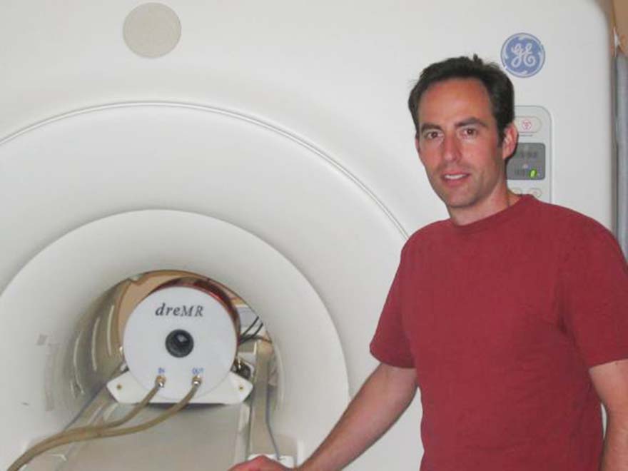
We have developed a novel shielded B0-shifting solenoid magnet that can be inserted into a clinical MR imager. This system is capable of field shifts of over 0.25T and has dedicated RF hardware capable of imaging small rodents. A fourth-gradient axis control system has developed and added as a slave channel to the clinical MR scanner to control the magnetic field of the shield insert magnet.
Research demonstrating dreMr-derived contrast using phantoms has been published as well as an article outlining hardware construction and other ancillary considerations.
Publications
-
J.K. Alford, T.J. Scholl, W.B. Handler, B.A. Chronik 2009, Design, construction, interfacing and benchmarking of a prototype high-power B0 insert coil for field-cycled imaging in a 1.5T superconducting MRI system, Concepts in Magnetic Resonance B: Magnetic Resonance Engineering 35B, 1-10.[PDF]
-
J.K. Alford, B.K. Rutt, T.J. Scholl, W.B. Handler, B.A. Chronik 2009, Delta relaxation enhanced MR: obtaining order of magnitude increases in the activation-specificity of molecular probes through R1 dispersion imaging, Magnetic Resonance in Medicine 61, 796-802.[PDF]
Key Questions
- What will be the expected SNR for dreMR contrast using new optimized double inversion pulse sequences?
- Will the enhanced specificity compensate for the reduction in SNR compared to ordinary contrast-enhanced MR?


